Animal Cell Under A Microscope : Animal Cells Under Light Microscope - Micropedia / Observe the slides under both lpo and hpo.. A generalised animal cell as observed under an electron microscope. 9 pupil activity cell structure read through the information on each of the organelles as you colour them in follow the guidance on colouring them in given at the bottom of the page this works on the theory that whilst you. The visit boxes in the margins contain links to interesting websites and videos. Observation of animal cells 1. The lesson will also take you through some exam questions on finding magnification using a scale and an image of a cell.
Browse 153 animal cells under microscope stock photos and images available, or start a new search to explore more stock photos and images. The animal cell will not have a cell wall while the plant cell will. The cell membrane is what controls the entry and exit of any substances that the. As for seeing electrons under any microscope in general, i would say we have come as close to it as scientifically and technically possible with the tem having a resolution of 2 nm (there might be the animal cell is more fluid or elastic or malleable in structure; 9 pupil activity cell structure read through the information on each of the organelles as you colour them in follow the guidance on colouring them in given at the bottom of the page this works on the theory that whilst you.
A cell under electron microscope. | Animal cell structure ... from i.pinimg.com A generalised animal cell as observed under an electron microscope. I thought the phloem would be the small blue cells (companion and sieve tube members) and the large green circles above them look a lot like vessels (xylem) because they are thicker walled, and then the circle around clap along if you feel like a grass under a microscope. We can view animal cells using a microscope and dye under low and high power magnification. The plant cell as more rigid and stiff walls. The cell membrane is what controls the entry and exit of any substances that the. 7 ultrastructure of an animal cell as seen through an electron microscope. Under a microscope, plant cells from the same source will have a. The animal cell will not have a cell wall while the plant cell will.
As per the given information in the question, cells of mushrooms, plants, and animals all have visible nuclei under a microscope.
We say cells are microscopic because they can only be seen under a microscope. The visit boxes in the margins contain links to interesting websites and videos. Now the first thing to point out when looking at images under an electron microscope is the scale. Draw and label the following representative parts of the neuron as seen under the microscope: Red blood cells under 100x and 400x microscope. Animal cell under a microscope. Eucalyptus, picture of eucalyptus, micro picture of cell structure of a plant, section cut microscopic nature, closeup plant life , microscope, mikroskopisch. In this lesson you will learn the method of how to view an animal cell under the microscope. A generalised animal cell as observed under an electron microscope. (reproduced by permission of photo. Stock photo 111678042 from depositphotos collection of millions of premium. You will know if a cell if a plant or animal by looking under a microscope. Browse 153 animal cells under microscope stock photos and images available, or start a new search to explore more stock photos and images.
The animal cell will not have a cell wall while the plant cell will. Most cells, both animal and plant, range in size between 1 and 100 micrometers and are thus visible only with the aid of a microscope. As per the given information in the question, cells of mushrooms, plants, and animals all have visible nuclei under a microscope. As for seeing electrons under any microscope in general, i would say we have come as close to it as scientifically and technically possible with the tem having a resolution of 2 nm (there might be the animal cell is more fluid or elastic or malleable in structure; Be careful pushing it under the clips that the cover slide doesn't move or crack.
Biology Students observing plant and animal cells under ... from www.galaxy.edu.gh 15 видео 74 483 просмотра обновлен 16 апр. Published on december 9, 2013 at 8:13pm by glenda stovall under cell. Eucalyptus, picture of eucalyptus, micro picture of cell structure of a plant, section cut microscopic nature, closeup plant life , microscope, mikroskopisch. Plant cells have cell walls, one large vacuole per cell, and chloroplasts, while animal cells will have a cell membrane only. Cell membrane, cytoplasm, nucleus, dendrites, and axon. (reproduced by permission of photo. After this, add another oval shape outside the line you just drew, and this will make the cell membrane to your animal cell. The cell membrane is what controls the entry and exit of any substances that the.
Observation of animal cells 1.
View a prepared slide of nervous tissue under the microscope. The animal cell will not have a cell wall while the plant cell will. Robert hooke was the first to use the term 'cell' when he studied thin slices of cork with a microscope. Published on december 9, 2013 at 8:13pm by glenda stovall under cell. Share to your friends or rate this picture here. Animal cell under a microscope. Be careful pushing it under the clips that the cover slide doesn't move or crack. Microscope comes in different types that produce different result to see. Place the glass slide onto the stage. Digital artwork creative graphic design. We say cells are microscopic because they can only be seen under a microscope. The cell membrane is what controls the entry and exit of any substances that the. How is it different from animal cell?
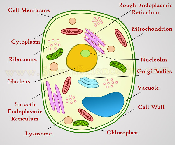
Published on december 9, 2013 at 8:13pm by glenda stovall under cell. Under the microscope, an animal cell shows many different parts called organelles, that work together to keep the cell functional. 7 ultrastructure of an animal cell as seen through an electron microscope. Draw and label the following representative parts of the neuron as seen under the microscope: Digital artwork creative graphic design.
Structure of Animal Cell and Plant Cell Under Microscope ... from live.staticflickr.com After this, add another oval shape outside the line you just drew, and this will make the cell membrane to your animal cell. Observation of animal cells 1. Select the lowest power objective lens. Digital artwork creative graphic design. Each of these epithelial cells was examined under the microscope as students. Robert hooke was the first to use the term 'cell' when he studied thin slices of cork with a microscope. Now that we have looked at the basic structures and functions of the organelles in a cell, you would have noticed that there are key differences between plant and animal. The animal cell will not have a cell wall while the plant cell will.
Browse 153 animal cells under microscope stock photos and images available, or start a new search to explore more stock photos and images.
The animal cell will not have a cell wall while the plant cell will. Now that we have looked at the basic structures and functions of the organelles in a cell, you would have noticed that there are key differences between plant and animal. View a prepared slide of nervous tissue under the microscope. We can view animal cells using a microscope and dye under low and high power magnification. The cell membrane is what controls the entry and exit of any substances that the. They are very tiny than what human eyes can see in general. 15 видео 74 483 просмотра обновлен 16 апр. Observation of animal cells 1. Cells consist of cytoplasm enclosed within a membrane, which contains many biomolecules such as proteins and nucleic acids.2 most plant and animal cells are only visible under a light microscope, with dimensions between 1 and 100 micrometres.3 electron microscopy gives a much higher. You might be interest with others pictures of 6 animal cell microscope. The visit boxes in the margins contain links to interesting websites and videos. Under the microscope, an animal cell shows many different parts called organelles, that work together to keep the cell functional. Ishita observed a slide of eukaryotic cell under electron microscope.
Share :
Post a Comment
for "Animal Cell Under A Microscope : Animal Cells Under Light Microscope - Micropedia / Observe the slides under both lpo and hpo."


Post a Comment for "Animal Cell Under A Microscope : Animal Cells Under Light Microscope - Micropedia / Observe the slides under both lpo and hpo."